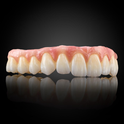
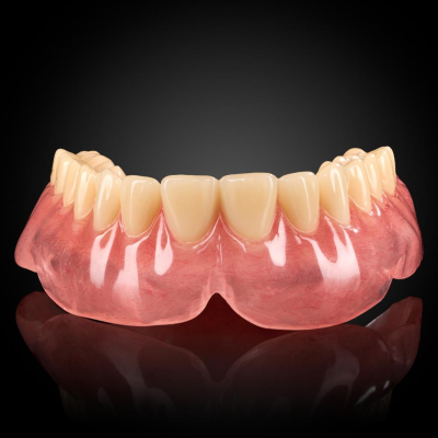
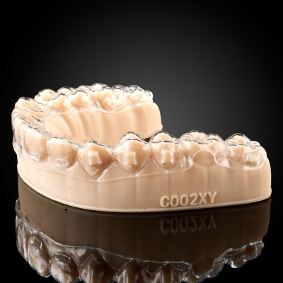
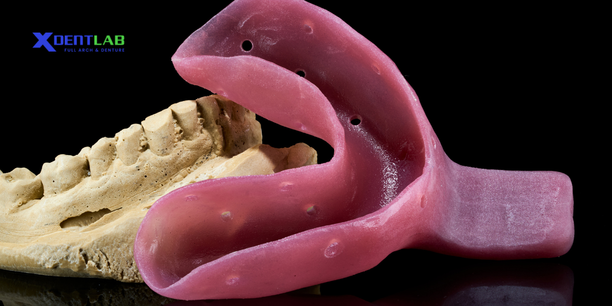
 Border of the Custom Tray for Full Dentures[/caption]
 Table of contents [Show]
The Principle of Retention in Dentures
The retention of a full denture relies on creating an airtight seal between the denture base and the patient’s mucosal surface. Placing two glass plates together with a drop of water in between easily demonstrates this principle. The cohesive properties of the water cause the plates to stick tightly, making them difficult to separate. In the same way, saliva acts as a natural adhesive between the denture and the oral tissues, ensuring the denture stays in place.Principle of Adhesion
Adhesion refers to the bonding force between the denture base and the mucosal surface, facilitated by a thin layer of saliva present between them.Mechanism of Action
Saliva forms a thin film between the denture base and the gingival tissue, enhancing frictional forces and reducing movement. The viscosity and quantity of saliva play a crucial role in this process.Factors Affecting Adhesion
- Surface Area of the Denture Base: A larger surface area results in stronger adhesion forces.
- Condition of Saliva: Thick and sufficient saliva improves adhesive strength.
The Role of the Vestibular Depth
The vestibular depth—the area where the fixed mucosa transitions into the movable mucosa—is crucial for maintaining this seal. If air enters the seal, the retention compromise, and the denture may become dislodged. Capturing the exact shape of the vestibular depth is therefore essential for a well-fitting denture.Challenges in Capturing the Vestibular Depth
One of the main difficulties in taking an accurate impression of the vestibular depth is its mobility. The movable mucosa tends to shift during the impression process, which can lead to inaccuracies. To overcome this, a specific technique is employed in dental labs during the tray design phase.Why the Tray Borders Are Positioned 1.5–2 mm Away
Custom trays are designed with their borders positioned 1.5–2 mm short of the vestibular depth to leave space for border molding wax, also known as Patondeker wax. This wax is applied to the tray’s edges and softened under heat to flow precisely along the vestibular contours. By doing so, the wax captures the exact shape of the patient’s oral anatomy, including the transitional zone between fixed and movable mucosa.Benefits of Proper Tray Border Design
At a professional dental lab, this meticulous approach offers several advantages:- Precision: Accurate impressions lead to a well-fitting final denture.
- Comfort: Properly fitting dentures reduce patient discomfort and enhance usability.
- Functionality: An accurate seal ensures the denture stays securely in place during daily activities.
Final Thoughts
The art of creating high-quality dentures involves a deep understanding of oral anatomy and precise fabrication techniques. By positioning the borders of a full-arch custom tray 1.5–2 mm away from the vestibular depth, dental labs ensure that every detail contributes to the retention, comfort, and effectiveness of the final product. [caption id="attachment_4772" align="aligncenter" width="1020"]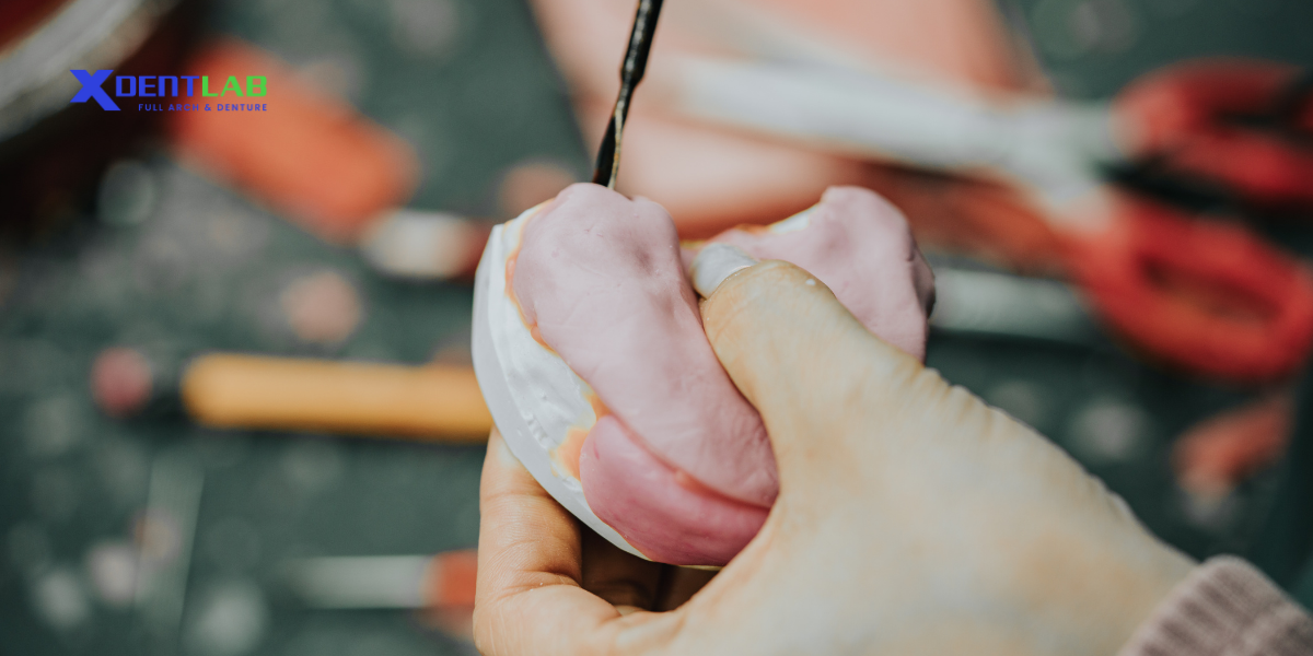 Dailywork in XDENT LAB[/caption]

