
Learn the anatomy and physiology of the floor of the mouth, including muscles, glands, functions, clinical significance, and its role in dental treatment planning.
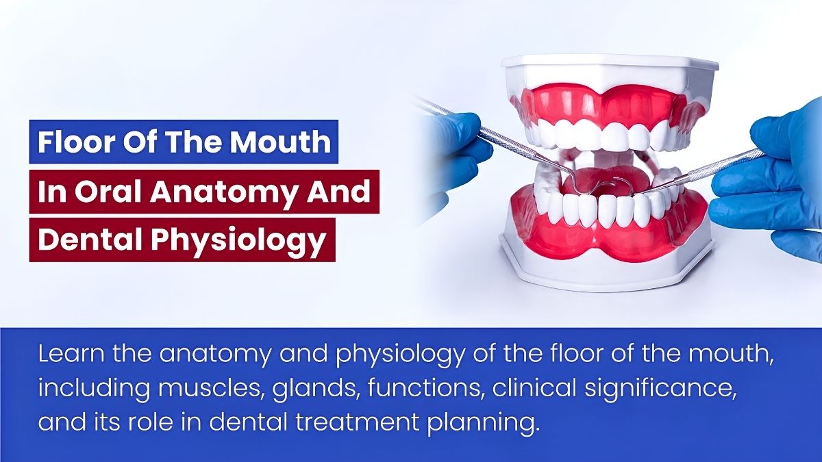
Table of contents [Show]
- What is the floor of the mouth?
- Anatomy of the floor of the mouth
- Physiology of the floor of the mouth
- Clinical significance in dentistry
- Common conditions affecting the floor of the mouth
- Diagnostic assessment of the floor of the mouth
- Importance for dental laboratory communication
- Emerging research and future directions
- Why this topic matters for dental practices and outsourcing labs
- Key takeaways
The floor of the mouth is a small anatomical region with outsized importance in oral health, speech, swallowing, salivary flow, and clinical dentistry. Located beneath the tongue and above the mylohyoid muscle layer, it contains soft tissues, muscles, ducts, glands, vessels, and nerves that work together to support daily oral function. For dental professionals, this area matters not only because of its anatomy, but also because it is involved in examination, diagnosis, local anesthesia, oral surgery, prosthodontic considerations, and pathology detection.
In dental physiology, the floor of the mouth contributes to lubrication, oral clearance, tongue mobility, and the maintenance of a stable environment for mastication and swallowing. In oral anatomy, it serves as a key landmark-rich region that supports the tongue and houses important salivary structures. This article explains the anatomy of the floor of the mouth, its physiological role, clinical significance, related disorders, diagnostic relevance, and why detailed anatomical understanding supports better dental outcomes across both conventional and digital workflows.
What is the floor of the mouth?
The floor of the mouth is the soft tissue region that forms the inferior boundary of the oral cavity proper. It lies under the tongue, inside the mandibular arch, and above the muscular sling primarily formed by the mylohyoid muscles.
Basic definition
From an anatomical perspective, the floor of the mouth is not a single structure. It is a complex region made up of:
- Mucosa
- Connective tissue
- Muscular layers
- Salivary glands
- Salivary ducts
- Blood vessels
- Nerves
- Fascial spaces
General location
It is located:
- Beneath the tongue
- Medial to the mandible
- Superior to the mylohyoid muscle
- Near the sublingual and submandibular salivary systems
This region is closely related to several oral and submandibular spaces, which is why infections and other pathologies can spread quickly if not diagnosed early.
Anatomy of the floor of the mouth
The anatomy of the floor of the mouth is clinically important because many essential oral functions depend on its structural relationships.
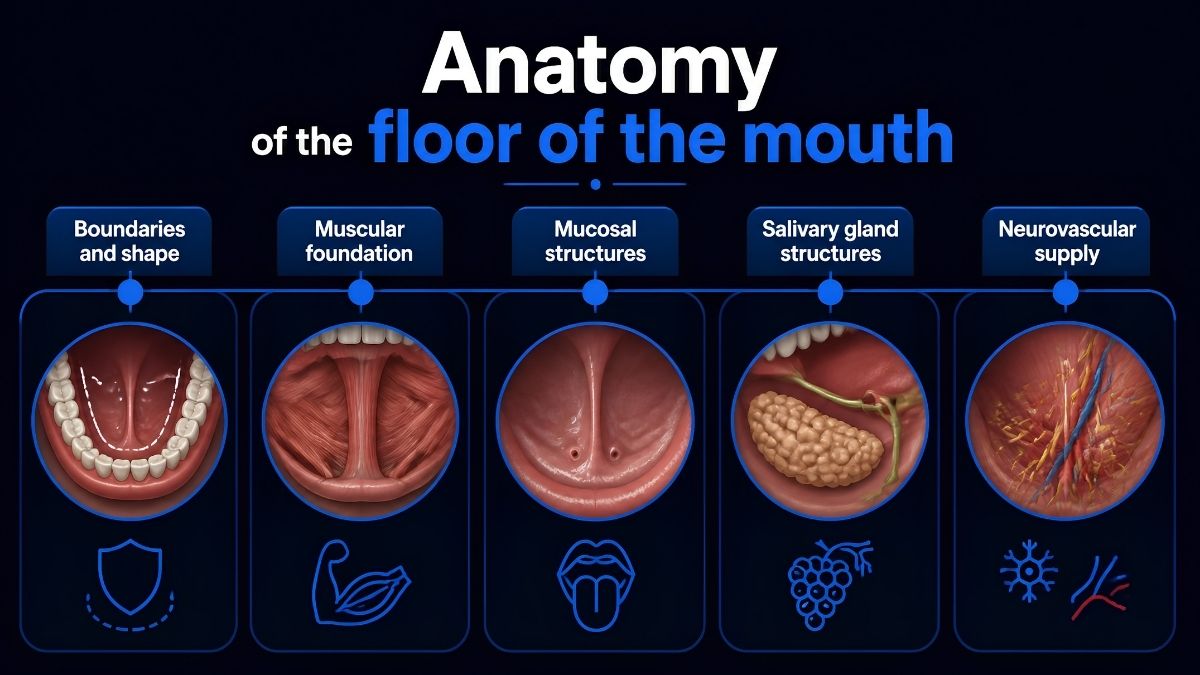
Boundaries and shape
The floor of the mouth is often described as a horseshoe-shaped area.
Main anatomical boundaries
Its boundaries include:
- The inner surface of the mandibular body and alveolar ridge
- The ventral surface of the tongue
- The mucosal covering over the muscular floor
- The posterior transition toward the oropharyngeal region
These relationships are highly relevant during intraoral examination, prosthetic planning, and surgery.
Muscular foundation
The muscular base of the floor of the mouth is primarily formed by the mylohyoid muscles, with important contribution from the geniohyoid muscles.
Mylohyoid muscle
The mylohyoid is the principal muscular diaphragm of the floor of the mouth.
Its main roles include:
- Supporting the floor of the oral cavity
- Elevating the floor during swallowing
- Assisting tongue function
- Helping stabilize oral soft tissues
Geniohyoid muscle
The geniohyoid lies superior to the mylohyoid and contributes to mandibular and hyoid movement.
Its functions include:
- Assisting in swallowing
- Helping move the hyoid bone anteriorly and superiorly
- Supporting coordinated tongue and floor movement
Together, these muscles create a dynamic platform rather than a static anatomical base.
Mucosal structures
The mucosa of the floor of the mouth is thin, delicate, and highly vascular.
Lingual frenum
The lingual frenum is a midline fold of mucous membrane connecting the underside of the tongue to the floor of the mouth.
It is clinically important because:
- It influences tongue mobility
- It may be associated with ankyloglossia
- It is a visible landmark during oral examination
Sublingual folds
The sublingual folds are raised ridges on either side of the lingual frenum.
They are formed by the underlying sublingual glands and contain multiple small duct openings.
Sublingual caruncles
The sublingual caruncles are small elevations located near the base of the lingual frenum.
They mark the openings of the submandibular ducts and are important landmarks in salivary assessment.
Salivary gland structures
The floor of the mouth contains major salivary components that are essential to oral physiology.
Sublingual glands
The sublingual glands are paired major salivary glands located beneath the mucosa of the floor of the mouth.
They primarily secrete:
- Mucous-rich saliva
- Lubricating secretions that help maintain oral moisture
Submandibular ducts
The submandibular glands themselves are mostly located below the mandible, but their ducts pass forward and open into the floor of the mouth.
These ducts:
- Transport saliva from the submandibular glands
- Open at the sublingual caruncles
- Play a major role in unstimulated salivary flow
Neurovascular supply
The floor of the mouth contains important nerves and blood vessels.
Key nerves
Relevant neural structures may include:
- Lingual nerve
- Hypoglossal nerve in deeper anatomical relation
- Nerve fibers associated with salivary gland function
Blood supply
The vascular supply comes primarily from branches of the lingual and facial arterial systems, with corresponding venous drainage.
This rich vascularity explains why the area is sensitive, heals relatively well, and may absorb medications quickly in some circumstances.
Physiology of the floor of the mouth
The floor of the mouth supports several essential physiological processes in the oral cavity.
Saliva secretion and distribution
One of the most important physiological functions of the floor of the mouth is its relationship to salivary production and release.
Why saliva matters
Saliva helps:
- Lubricate oral tissues
- Begin digestion
- Facilitate swallowing
- Protect teeth from demineralization
- Buffer oral acids
- Support soft tissue health
- Cleanse the oral cavity
Because the sublingual and submandibular systems open into this area, the floor of the mouth is central to oral moisture regulation.
Tongue mobility and coordination
The floor of the mouth provides structural and muscular support for tongue movement.
Functional relevance
This support is necessary for:
- Speech articulation
- Food manipulation
- Bolus formation
- Swallowing
- Oral clearance
Any restriction, inflammation, or surgical change in this area can affect oral function significantly.
Swallowing mechanics
The coordinated elevation of the floor of the mouth during deglutition is essential for normal swallowing.
During swallowing
The muscles of this region help:
- Elevate the tongue and floor
- Move the bolus posteriorly
- Coordinate with suprahyoid structures
- Support safe oral-to-pharyngeal transfer
Oral homeostasis
The floor of the mouth contributes indirectly to the maintenance of the oral environment.
Physiological contributions
Through its salivary relationships, it supports:
- pH balance
- Mucosal hydration
- Mechanical cleansing
- Antimicrobial defense
- Patient comfort
This is especially relevant in patients with xerostomia, salivary dysfunction, or systemic conditions that alter oral physiology.
Clinical significance in dentistry
For dentists and dental laboratories, the floor of the mouth is more than an anatomical description. It is a practical clinical region that affects diagnosis, treatment planning, impression making, prosthodontics, surgery, and patient safety.
Oral examination and early detection
The floor of the mouth is a high-priority region during routine intraoral examination.
Why careful examination matters
This area should be assessed for:
- Swelling
- Ulceration
- Color changes
- Mucosal lesions
- Salivary flow abnormalities
- Tenderness
- Masses or asymmetry
Because some oral malignancies and salivary pathologies may arise here, systematic examination is essential.
Relevance for local anesthesia
The floor of the mouth has close anatomical relationships with structures involved in intraoral injection techniques.
Clinical importance
Precise anatomical knowledge helps reduce the risk of:
- Nerve injury
- Hematoma
- Inadequate anesthesia
- Soft tissue trauma
The thin mucosa and vascularity of the area also make technique sensitivity especially important.
Although implant fixtures are not placed in the floor of the mouth itself, the region is highly relevant to surgical planning in the mandible.
Surgical considerations
Clinicians must understand:
- Soft tissue anatomy
- Lingual undercut risk
- Vascular considerations
- Floor elevation and swelling risk
- Potential spread of infection
This is particularly important in posterior mandibular surgery, flap management, and management of surgical complications.
Prosthodontic relevance
The anatomy of the floor of the mouth influences the extension and adaptation of removable prostheses, especially mandibular appliances.
Why it matters for removable cases
The functional depth and movement of the floor of the mouth can affect:
- Lingual flange design
- Major connector selection
- Patient comfort
- Denture stability
- Border molding accuracy
For dental labs working on removable prosthetics, accurate capture of these anatomical dynamics supports better-fitting restorations.
Impression and digital scanning implications
The mobility of the floor of the mouth can influence how intraoral data is captured.
In conventional and digital workflows
This area may affect:
- Soft tissue displacement
- Border extension accuracy
- Scan completeness
- Case communication between clinic and lab
For outsourcing dental labs, understanding these anatomical variables helps interpret clinical records more accurately.
Common conditions affecting the floor of the mouth
Several oral and maxillofacial conditions may involve the floor of the mouth.
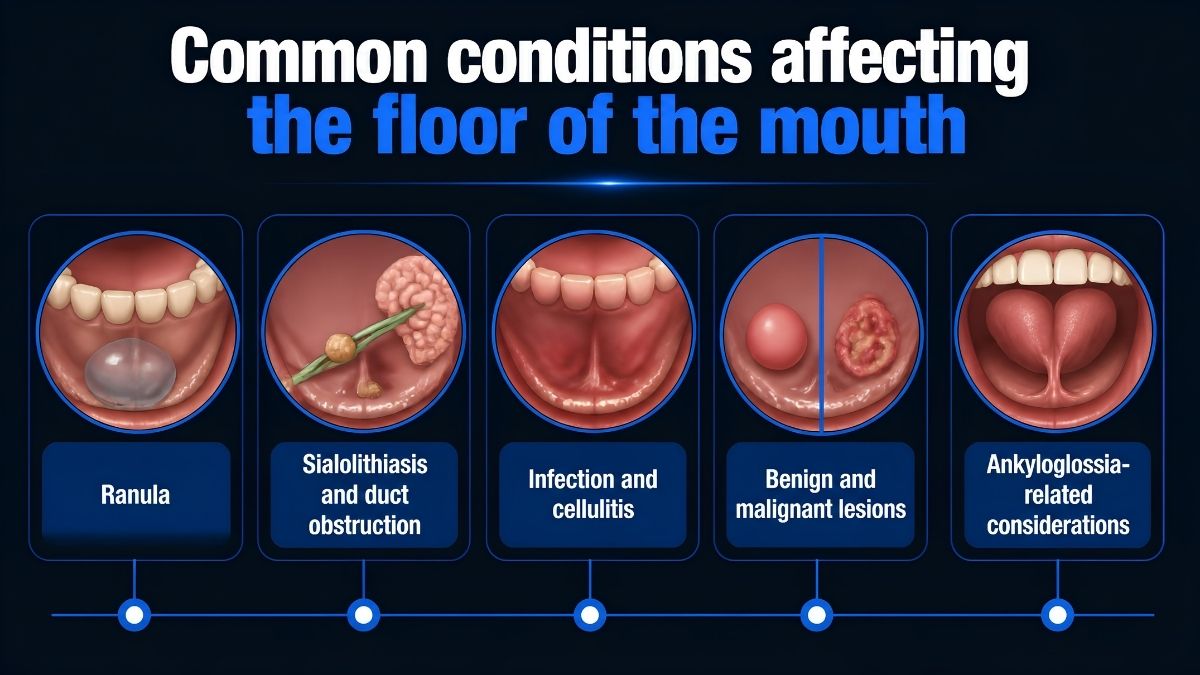
Ranula
A ranula is a mucus extravasation cyst associated with the sublingual gland.
Typical features
It may appear as:
- A bluish, fluctuant swelling
- A unilateral lesion in the floor of the mouth
- A lesion that interferes with speech or swallowing if large
Sialolithiasis and duct obstruction
The submandibular duct may become obstructed by salivary stones.
Clinical signs
Patients may present with:
- Pain during meals
- Swelling in the floor of the mouth
- Reduced salivary flow
- Recurrent gland inflammation
Infection and cellulitis
Infections involving the floor of the mouth may spread into adjacent fascial spaces.
Why this matters
Severe infections can compromise:
- Swallowing
- Airway safety
- Systemic health
This makes rapid diagnosis and referral essential.
Benign and malignant lesions
The floor of the mouth may be a site for cysts, leukoplakic changes, salivary lesions, and oral squamous cell carcinoma.
Clinical warning signs
Suspicious findings may include:
- Persistent ulceration
- Induration
- Non-healing lesions
- Erythroplakia or leukoplakia
- Unexplained swelling
- Pain or paresthesia
Although primarily a lingual frenum condition, ankyloglossia directly involves the floor of the mouth region.
Functional effects
It may contribute to:
- Restricted tongue movement
- Speech difficulty
- Swallowing issues
- Oral hygiene challenges in selected cases
Diagnostic assessment of the floor of the mouth
Proper assessment combines visual inspection, palpation, functional observation, and when needed, imaging.
Clinical examination approach
Routine examination should be systematic.
Basic steps
Clinicians often assess:
- Mucosal appearance
- Symmetry
- Tongue elevation and mobility
- Salivary flow from duct openings
- Palpable masses
- Tenderness or induration
Bimanual palpation is especially useful for evaluating glandular and soft tissue abnormalities.
Imaging and adjunctive evaluation
When pathology is suspected, imaging may support diagnosis.
Common diagnostic tools
Depending on the case, evaluation may involve:
- Ultrasound
- CBCT in selected dental contexts
- MRI for soft tissue characterization
- CT for infection spread or deep lesions
- Sialography in selected salivary assessments
Advanced imaging is increasingly helpful in differentiating soft tissue, glandular, and space-related pathology.
Importance for dental laboratory communication
Even though the floor of the mouth is primarily a clinical anatomical region, its relevance extends into laboratory communication and prosthetic manufacturing.
For removable prosthetics
Mandibular removable prostheses are strongly influenced by soft tissue anatomy and function.
A dental lab must understand how floor-of-mouth anatomy affects:
- Lingual extension design
- Relief requirements
- Connector selection
- Functional fit
- Patient adaptation
For digital workflows
Digital records are only as useful as the anatomical accuracy behind them.
Why lab communication matters
When dentists communicate details about tissue mobility, flange limitations, or special anatomy, the lab can produce restorations with better functional outcomes.
For a full-service outsourcing partner such as XDENT LAB, this type of anatomical awareness supports more accurate removable fabrication and stronger collaboration with dental practices seeking consistency and quality.
Emerging research and future directions
The floor of the mouth remains relevant in both clinical research and applied dental innovation.
Imaging and diagnostic improvement
New imaging approaches continue to improve early detection of lesions and more detailed assessment of soft tissues and salivary structures.
Salivary gland science
As xerostomia, systemic disease, and medication-related salivary dysfunction become more common, the study of sublingual and submandibular gland function will remain important.
Regenerative and reconstructive approaches
Future developments may include improved methods for:
- Soft tissue repair
- Salivary gland regeneration
- Post-surgical functional rehabilitation
- Better management of trauma and pathology-related tissue loss
Why this topic matters for dental practices and outsourcing labs
A strong understanding of oral anatomy improves every stage of dental care, from diagnosis to restorative delivery. The floor of the mouth is especially important because it intersects anatomy, function, pathology, prosthodontics, and surgical safety.
For dental practices, this knowledge supports better clinical examination, treatment planning, impression accuracy, and patient outcomes. For dental lab partners, especially those involved in removable prosthetics and complex case support, understanding the functional anatomy of this region improves communication and restorative precision.
XDENT LAB’s focus on removable and implant workflows makes this type of anatomical literacy especially relevant. In lab-to-lab outsourcing, precision is not only about manufacturing. It also depends on understanding how oral structures behave in real clinical conditions.
Key takeaways
The floor of the mouth is a vital anatomical and physiological region beneath the tongue that supports saliva secretion, tongue mobility, swallowing, and oral homeostasis. It contains important muscles, glands, ducts, mucosal landmarks, nerves, and blood vessels that make it highly relevant to clinical dentistry.
Its significance extends beyond anatomy into oral examination, pathology detection, anesthesia, surgery, removable prosthodontics, and dental lab communication. For practices and outsourcing partners alike, a detailed understanding of the floor of the mouth contributes to safer procedures, better prosthetic design, and more predictable dental outcomes.
About XDENT LAB:
We are experts in Lab-to-Lab Full Service from Vietnam, with the signature services of Removable, meet U.S. market standards, FDA-registered, ISO 13485-certified. Founded in 2017, from local root to global reach, we scale with 2 Factories with over 100+ employees.

Our Commitments Built on “Trusted. Commitment. Quality”
- Commit to Large-Scale Manufacturing, high volume, remake rate < 1%.
- Commit to 5-Year Warranty
- Commit to Competitive Price
XDENT LAB | A Trusted Lab-to-Lab Service from Vietnam
Share this post:
Related Posts

Learn how vestibular depth in dentistry affects oral anatomy, denture retention, implant planning, periodontal health, clinical assessment, and treatment decisions.
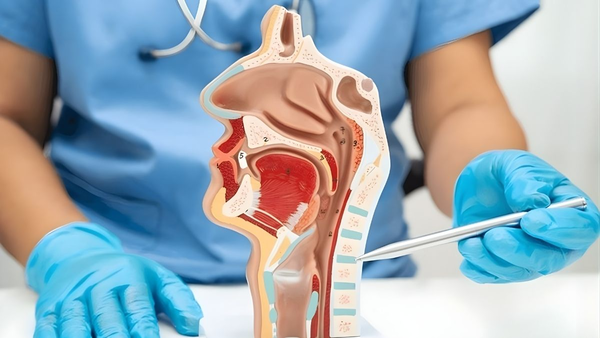
Explore oral cavity anatomy and function, including key structures, roles in speech and digestion, clinical relevance, and why it matters for dental professionals.

