
Explore maxillofacial prosthodontics, from obturators and facial prostheses to materials, digital design and dental lab workflows.
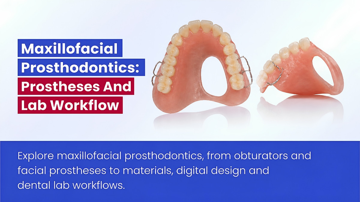
Table of contents [Show]
Maxillofacial prosthodontics helps people whose oral or facial structures are missing, altered or unable to function normally. Treatment may follow cancer surgery, trauma or a congenital condition, or address difficulties caused by neuromuscular disease. The goal is not simply to reproduce anatomy: a prosthesis should help the patient speak, swallow, eat, look and participate in daily life as comfortably as possible.
For dental practices and laboratories, these cases require unusually close coordination. A successful device depends on the clinical defect, remaining tissues, retention, material suitability, patient handling and a plan for adjustment or replacement. This guide covers the broader specialty—from obturators and facial prostheses to implant-assisted rehabilitation, digital workflows and quality control.
What does maxillofacial prosthodontics include?
The American Academy of Maxillofacial Prosthetics describes maxillofacial prosthetics as a subspecialty of prosthodontics concerned with congenital and acquired defects or disabilities. Its scope includes oral function, facial rehabilitation and selected appliances used during radiation treatment.
Who is involved in treatment?
Depending on the case, the team may include a maxillofacial prosthodontist, surgeon, oncologist, speech-language pathologist, swallowing specialist, dental technician, anaplastologist, ocularist and other rehabilitation professionals. An anaplastologist specializes in restoring external anatomy; an ocularist specializes in artificial eyes. Professional roles and qualifications vary by jurisdiction.
Where circumstances permit, prosthodontic planning should begin before surgery or radiotherapy. Early discussion can help preserve useful structures, anticipate the eventual insertion path, plan implants when appropriate and establish realistic functional goals. Prosthetic rehabilitation and surgery are often complementary—not competing—approaches.
What types of prostheses are used?
Devices are best distinguished by the problem they address. One patient may require more than one type during rehabilitation.
| Prosthesis or appliance | Typical purpose | Important distinction |
|---|---|---|
| Surgical, interim or definitive obturator | Closes an oral–nasal or related maxillary defect | Design changes as tissues heal |
| Speech bulb | Assists closure when velopharyngeal tissue is structurally insufficient | Requires functional speech assessment |
| Palatal lift | Elevates a soft palate with inadequate movement | Addresses movement rather than simply replacing missing tissue |
| Palatal augmentation prosthesis | Reshapes the palate to improve contact with a reduced or impaired tongue | Contour is guided by speech and swallowing function |
| Mandibular guidance appliance | Helps guide jaw position after selected resections | Does not guarantee correction of deviation |
| Auricular, nasal, orbital or midfacial prosthesis | Replaces missing external facial anatomy | Margins, color and daily handling are central to success |
| Ocular prosthesis | Restores the appearance of an eye within a suitable socket | Does not restore vision |
| Oncology-related appliance | Supports a specified treatment or oral-care protocol | Requires direction from the relevant clinical team |
These categories overlap in practice. For example, a patient may receive an obturator during healing, implant-assisted dental rehabilitation later and a separate facial prosthesis for an external defect.
How are maxillary obturators planned?
An obturator closes a communication created by a maxillary defect. It may also replace teeth or support surrounding contours. Treatment often progresses through three stages:
- Surgical obturator: Planned before surgery when feasible and used according to the surgical team’s protocol.
- Interim obturator: Adjusted or relined as swelling resolves and tissues change.
- Definitive obturator: Fabricated when the defect and wider treatment situation are sufficiently stable.
Design depends on the size and location of the defect, remaining teeth, tissue tolerance, available undercuts, mouth opening and the patient’s ability to insert and clean the device. A hollow bulb may reduce weight, but a closed bulb needs a dependable seal. An open bulb presents different cleaning and fluid-management considerations. Neither is universally preferable.
Laboratory priority: Reduce unnecessary bulk without creating a fragile, porous or inaccessible prosthesis. Speech, swallowing and nasal leakage must be assessed clinically; an apparently close-fitting model does not prove functional sealing.
How do speech, palatal and mandibular appliances differ?
A speech bulb helps compensate for insufficient tissue at the velopharyngeal opening. A palatal lift elevates tissue that is present but moves inadequately. A palatal augmentation prosthesis changes the palatal surface so a reduced or less mobile tongue can make more effective contact. Their shapes should be refined with functional assessment, often involving a speech-language pathologist.
After some mandibular resections, a guide flange or palatal guidance ramp may assist jaw positioning. The result depends on the extent of resection, remaining movement, scarring and rehabilitation. For a reconstructed jaw, treatment also requires coordination of bone position, implant trajectory, soft-tissue thickness, tongue space, occlusion and cleaning access.
A systematic review of dental implant rehabilitation after maxillofacial reconstruction examines patient quality of life. It reinforces the importance of considering dental function during reconstruction planning, while conclusions remain dependent on the quality and comparability of the underlying studies.
Materials and retention: Matching the device to its purpose
Maxillofacial devices can contact skin, mucosa, implants or several tissues at once. Material selection must therefore follow the specific intended use and contact conditions, not a general description such as “medical grade.”
Which materials are commonly used?
The principal options differ in flexibility, appearance, rigidity and repair methods.
| Material | Typical use | Key control |
|---|---|---|
| Maxillofacial silicone elastomer | External facial prostheses | Thin-edge strength, color, cure and skin-contact suitability |
| PMMA | Obturators, intraoral bases and rigid substructures | Fit, strength, finishing and repairability |
| Approved printed polymer | Models, patterns and selected definitive devices | Exact intended use and validated post-processing |
| Cobalt–chromium | Selected intraoral frameworks | Alloy specification, fit and finishing |
| Titanium | Implant components, bars and selected frameworks | Component compatibility and fit verification |
| Soft liner or tissue conditioner | Selected interim applications | Aging, hygiene and replacement interval |
A resin intended for printing a dental model is not automatically suitable for a definitive obturator or prolonged mucosal contact. Similarly, a pigment, primer or adhesive must be compatible with both the base material and its intended clinical application.
Why does facial silicone need careful processing?
Silicone can reproduce flexibility, translucency and surface texture, but its performance depends on the exact formulation and processing protocol. Thin margins may tear; color can change with exposure and cleaning; bonds to rigid substructures may fail.
The laboratory should control mixing ratios, pigment loading, de-airing, mold preparation, cure conditions and bonding procedures. Some silicone systems can be affected by contaminants that inhibit curing, so material compatibility must be checked for the product being used.
Color matching combines intrinsic pigmentation with surface characterization. Clinical review under more than one lighting condition is valuable because surrounding skin changes with lighting, temperature and expression. A systematic review of maxillofacial silicone color stability highlights the influence of formulation and exposure. Laboratory aging results, however, cannot be translated reliably into one universal clinical replacement interval.
How are facial prostheses retained?
Retention may come from anatomical undercuts, skin adhesives, spectacles, craniofacial implants or combinations of these methods. Implant-assisted bars, clips and magnets can improve repeatable positioning in selected cases, but they add cleaning and component-maintenance requirements.
The best method is one the patient can use consistently. Retention must be balanced against skin tolerance, dexterity, removal technique and the durability of thin silicone edges. Implants do not prevent silicone aging or eliminate the possibility of a future remake.
For previously irradiated patients, implant planning is site-specific. Radiation exposure, bone and soft-tissue condition, surgical feasibility and expected maintenance all matter. Neither a universal implant-survival figure nor a single “safe” radiation dose is appropriate for every maxillofacial case.
Conventional, digital and hybrid laboratory workflows
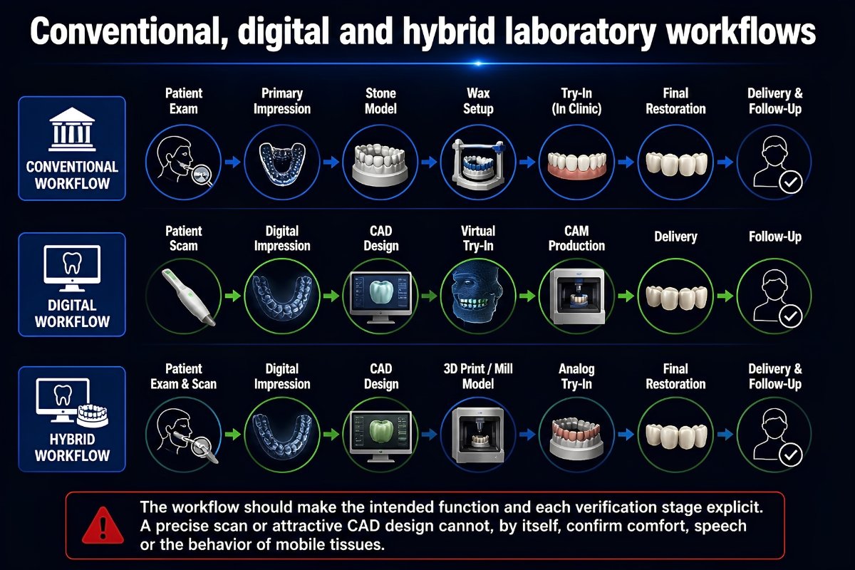
The workflow should make the intended function and each verification stage explicit. A precise scan or attractive CAD design cannot, by itself, confirm comfort, speech or the behavior of mobile tissues.
What information should accompany the prescription?
A useful clinical–laboratory handoff identifies:
- The defect, treatment stage and primary functional goals.
- Relevant surgery, planned radiotherapy and tissue sensitivities.
- Remaining teeth, implants and exact component identifiers.
- The proposed insertion path, retention method and contact tissues.
- Approved material systems and any intended-use restrictions.
- Records for tooth position, facial symmetry, color or speech assessment, as applicable.
- Who will verify the trial device, framework and final fit.
- Cleaning instructions, repair options and anticipated tissue changes.
Impressions involving deep defects, orbital areas or structures near the airway require appropriately trained clinical personnel and protective procedures. They are not routine laboratory-only tasks.
What happens in a conventional workflow?
After diagnostic planning, the clinician captures the anatomy using an appropriate impression or other record. The laboratory prepares a model and trial device, then checks contours, margins, insertion path and attachment positions with the treating team. Framework or substructure fit is verified before definitive processing when indicated.
For a facial prosthesis, mold fabrication, silicone processing, edge finishing and color characterization follow the approved trial. For an intraoral device, processing may instead involve acrylic, a metal framework or another validated construction. At delivery, the clinician assesses retention, pressure, function and the patient’s ability to manage the prosthesis.
Where does digital manufacturing help?
Facial scanning, intraoral scanning, imaging, photogrammetry and CAD can help document anatomy and plan a design. Digital mirroring of the unaffected side may provide a starting shape. Printed patterns or molds can then support conventional material processing.
It is important to distinguish two routes:
- Indirect digital manufacture: Capture and design digitally, then print a pattern or mold and process a suitable final material.
- Direct digital manufacture: Print the definitive prosthesis material itself.
A systematic literature review of intraoral maxillofacial digital workflows examines how much of the process has been digitized in published cases. A scoping review of directly printed soft maxillofacial prostheses maps emerging techniques. These publications support careful evaluation of digital tools; they do not establish that every directly printed soft prosthesis matches the long-term performance of a conventionally processed silicone device.
Deep defects, mobile tissues, facial expression and incomplete color capture remain practical challenges. A mirrored ear is a design reference, not a finished prosthesis: natural asymmetry, margin position and the patient’s preferences still require clinical judgment.
Which checks belong in the quality record?
Documentation should connect the approved design to the materials and steps used to produce it. Depending on the device, records may include material and implant-component lot numbers, pigment formulations, cure conditions, printing and post-curing settings, framework inspection, bonding protocols and clinical fit feedback.
Digital photographs, scans and imaging are sensitive patient records. Their use and storage should follow applicable consent, privacy and access-control requirements.
Clinical outcomes, maintenance and laboratory responsibility
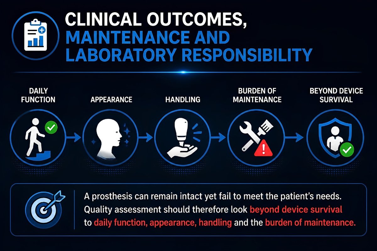
A prosthesis can remain intact yet fail to meet the patient’s needs. Quality assessment should therefore look beyond device survival to daily function, appearance, handling and the burden of maintenance.
Obturator or surgical reconstruction: Is one better?
These approaches solve different problems and are sometimes combined. An obturator can offer adjustability and access to the defect but requires insertion, cleaning and adequate retention. Surgical reconstruction restores tissue continuity but adds surgical demands and may still require later dental rehabilitation.
A systematic review and meta-analysis comparing obturation and reconstruction examines reported patient outcomes. Its findings should not be used to declare the methods interchangeable for every maxillary defect. Defect extent, remaining dentition, radiation history, general health and patient preference must guide selection.
What outcomes and complications matter most?
Clinical review may consider speech intelligibility, nasal leakage, swallowing, chewing, comfort, appearance satisfaction, cleaning difficulty and time spent handling the device. Technical records should distinguish repairs, relines, attachment replacement and complete remakes. Implant survival, prosthesis survival and a satisfactory patient outcome are not the same endpoint.
Common problems include obturator leakage as tissues change, fracture in thin intraoral sections, torn facial margins, silicone discoloration, attachment wear and inflammation around implants. Persistent poor speech may require a joint prosthodontic and speech assessment rather than another laboratory contour change alone.
In oncology patients, xerostomia, mucosal fragility, fibrosis and limited mouth opening can alter both fit and maintenance. New ulceration, unexplained bleeding, progressive pain or a sudden change in fit calls for clinical assessment, not merely a prosthesis adjustment. Radiation-positioning or shielding appliances must be planned with the radiation-oncology and medical-physics team; the laboratory should not independently infer dosimetric safety.
How do standards support quality assurance?
Depending on the device and material, relevant standards may include ISO 20795-1 for denture-base polymers, ISO 22674 for metallic dental materials, ISO 7405 and the ISO 10993 series for biological evaluation, ISO 13485 for medical-device quality management, and ISO 14971 for risk management. ISO 14801 concerns defined implant fatigue testing; it is not certification of an entire implant-retained facial or dental prosthesis. Current editions and scopes should be checked through the official ISO catalogue.
Material testing, quality-system controls and clinical suitability are distinct. Passing a mechanical test does not establish acceptable color stability, biological safety for every contact condition or success in a specific patient.
What is XDENT LAB’s role?
For dental practices using a Vietnam dental lab or dental lab outsourcing, complex maxillofacial cases need an especially clear division of responsibilities. The treating specialists establish the clinical objectives, perform sensitive anatomical capture, select treatment and verify the device in the patient. The laboratory manufactures to the approved prescription, documents its materials and processes, and flags fit, compatibility or serviceability concerns.
XDENT LAB’s lab-to-lab removable and implant experience can support the fabrication and documentation of suitable prescribed components. Capabilities, material indications and case requirements should be confirmed for each project. Clinical rehabilitation, including speech, oncology and surgical decisions, remains with the patient’s treating team.
Key takeaway: Maxillofacial prosthodontics works best when the prosthesis is planned as part of a broader rehabilitation—not as a stand-alone replacement for missing anatomy. Function, appearance, material safety, verifiable manufacture and a realistic maintenance plan all deserve attention from the start.
About XDENT LAB:
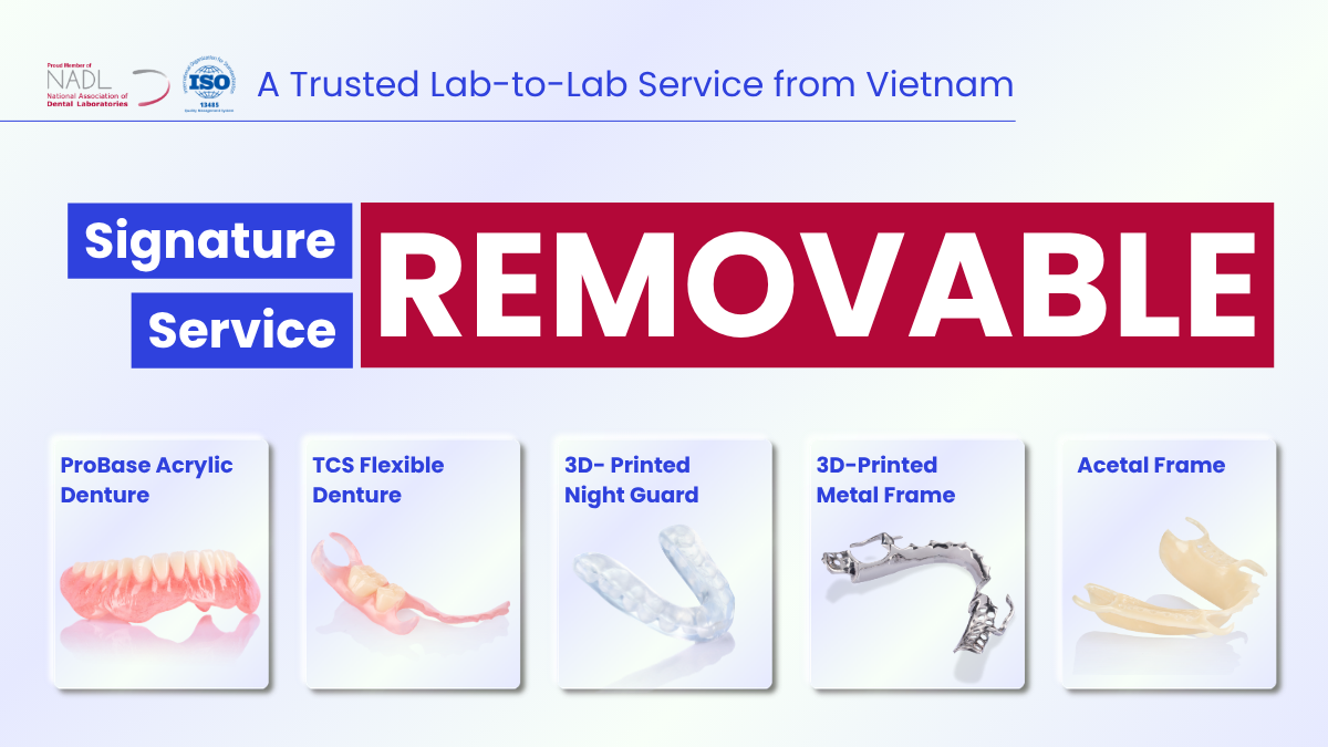
We are experts in Lab-to-Lab Full Service from Vietnam, with the signature services of Removable, meet U.S. market standards, FDA-registered, ISO 13485-certified. Founded in 2017, from local root to global reach, we scale with 2 Factories with over 100+ employees.
Our Commitments Built on “Trusted. Commitment. Quality”
- Commit to Large-Scale Manufacturing, high volume, remake rate < 1%.
- Commit to 5-Year Warranty
- Commit to Competitive Price
XDENT LAB | A Trusted Lab-to-Lab Service from Vietnam
Share this post:
Related Posts
A guide for dental practices to implant-supported overdenture planning, attachment selection, digital workflows and long-term care.
Explore full-arch fixed prosthesis planning and lab workflows, from implant distribution and framework fit to materials, hygiene and maintenance.
A practical guide to implant-supported bridges for dental practices, covering retention, materials, cleanable contours and lab quality checks.

