
A practical guide to implant-supported bridges for dental practices, covering retention, materials, cleanable contours and lab quality checks.
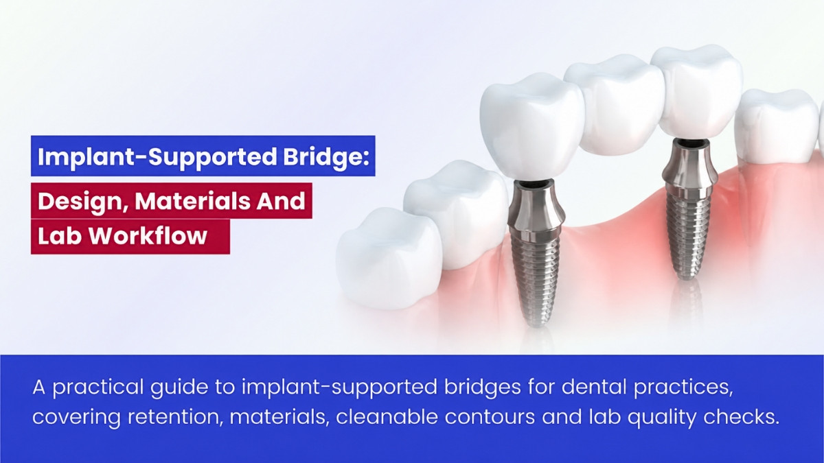
Table of contents [Show]
An implant-supported bridge replaces several missing teeth with a fixed restoration anchored to dental implants. The patient does not remove it, although many designs can be retrieved by a clinician for maintenance or repair.
A successful bridge depends on more than implant integration or a strong framework. Implant position, support distribution, component compatibility, fit, restorative material and cleaning access must work together. For dental practices and laboratories, it is best understood as a coordinated clinical–laboratory system—not simply several implant crowns joined together.
What is an implant-supported bridge?
In academic literature, an implant-supported bridge is often called an implant-supported fixed dental prosthesis, or FDP. Older publications may use the term fixed partial denture.
Components and common configurations
A bridge may include implants, abutments or other intermediate components, prosthetic screws, a framework or monolithic structure, and replacement teeth. A pontic is a replacement tooth without an implant directly beneath it.
| Configuration | Description | Key planning concern |
|---|---|---|
| Bounded-span bridge | One or more pontics between implant supports | Support distribution, connectors and cleaning beneath pontics |
| Cantilever bridge | A replacement unit extends beyond a terminal support | Bending forces, loading position and design limits |
| Splinted implant restorations | Adjacent implant-supported units joined together | Combined fit, access and load distribution |
| Full-arch fixed prosthesis | Restoration of an entire edentulous arch | Implant distribution, tissue replacement, speech and hygiene |
A three-unit bridge supported by two implants is one possible design, not a rule for every three-tooth space. Anatomy, loading, implant positions, restorative space and component indications determine whether a configuration is suitable.
A bridge connecting a natural tooth to an implant requires separate consideration. Teeth move slightly through their periodontal ligaments; implants have substantially less movement. Evidence for an entirely implant-supported bridge should not be applied to a tooth–implant-supported design without qualification.
When is an implant-supported bridge considered?
An implant bridge may be considered when several adjacent teeth are missing, appropriate implant positions are available and a fixed restoration suits the patient’s needs. Treatment planning should also compare preservation of restorable teeth, individual implant crowns, a conventional bridge and removable alternatives.
Implants are not automatically preferable to retaining restorable teeth. The decision should account for prognosis, biological cost, patient preference and expected lifetime maintenance.
Why should the restoration guide implant planning?
The proposed tooth arrangement should establish where the implants need to support the bridge. Diagnostic arrangements or digital wax-ups, scans, photographs, occlusal records and clinically justified imaging can connect the intended restoration to the available anatomy.
Before fabrication, the clinical team and laboratory should establish:
- Final tooth positions and available restorative space.
- Implant emergence and screw-access locations.
- Support distribution and any proposed cantilever.
- Occlusion and opposing dentition.
- Pontic contours and patient cleaning access.
- Retrieval, repair and maintenance plans.
A well-integrated implant in an unsuitable position can still make the bridge difficult to design or clean. Angulated components can help in selected situations, but they cannot resolve every consequence of poor implant positioning.
Implant number and cantilever design
There is no universal formula for the number of implants needed per replacement tooth. Span, implant distribution, crown height, material, opposing dentition and patient-specific loading all influence the plan.
Cantilevers increase bending demands, but length alone does not describe the entire risk. The laboratory should not add an unplanned cantilever without clinical approval and confirmation that the proposed components and materials support the design.
Retention methods and restorative materials
The choice of retention and material affects appearance, manufacturing, complication risks and future access. Neither the strongest material nor the most convenient attachment method is automatically the best choice.
Screw-retained versus cement-retained bridges
| Consideration | Screw-retained bridge | Cement-retained bridge |
|---|---|---|
| Retrieval | Generally more straightforward | May be difficult or require damage to the restoration |
| Intraoral cement | Avoided at bridge delivery | Excess must be minimized and removed |
| Access openings | Required through the restoration | Usually absent from the visible bridge surfaces |
| Potential complications | Screw loosening, fracture or access-filling loss | Loss of retention or cement-associated tissue concerns |
The ITI consensus resource on screw- and cement-retained fixed reconstructions provides a framework for evaluating these trade-offs. Both approaches can perform well, but neither removes the need to manage technical and biological complications.
For cement-retained bridges, margins should allow excess cement to be identified and removed. For screw-retained designs, access locations must allow effective tightening and future retrieval. Approved angulated screw channels require their specified screws, drivers and torque instructions.
A bridge bonded to titanium bases outside the mouth may still be screw-retained at the implants. This is different from cementing a bridge intraorally over abutments.
Implant-level and abutment-level connections
A bridge may connect directly to implant platforms or to intermediate abutments. The choice affects restorative space, insertion path, tissue interface and component selection.
Engaging and non-engaging components must be selected according to the implant system and the planned restoration. Components appropriate for a single implant crown should not automatically be substituted into a splinted bridge.
Materials used for implant-supported bridges
Each material family has different design rules and maintenance implications.
| Material or system | Potential advantage | Design consideration |
|---|---|---|
| Metal–ceramic | Established clinical use and a rigid framework | Veneer chipping and possible esthetic limitations |
| Monolithic zirconia | Avoids a separate veneering layer | Product-specific span, connector, thickness and finishing rules |
| Veneered zirconia | Allows individualized characterization | Additional veneer-related complication pathway |
| Titanium framework | Low density and corrosion resistance | Fit, joining and veneering or bonding compatibility |
| Cobalt–chromium framework | High stiffness | Manufacturing accuracy and appropriate finishing |
| Framework-supported acrylic or composite | Repair options for selected designs | Wear, discoloration and bond maintenance |
A material approved for a single crown is not necessarily approved for an implant bridge. In particular, bridge use of lithium disilicate or a given zirconia grade must be supported by the specific material and component manufacturers’ indications.
Connectors, pontics and hygiene contours
Connectors need product-appropriate dimensions and geometry to support the intended span. Screw channels, sharp transitions and inadequate connector sections can create critical areas in a bridge.
Pontics and tissue-facing surfaces must balance appearance with cleaning access. Excessive tissue pressure or inaccessible concavities can make routine hygiene difficult, particularly in full-arch designs. A polished surface is not automatically a cleanable surface if the patient cannot reach it.
Dental lab workflow and fit verification
Multi-implant restorations make accurate records especially important: components may appear to fit individually but fail to seat together as a bridge. Reliable fabrication therefore depends on verification throughout the clinical–laboratory workflow.
What should the laboratory prescription include?
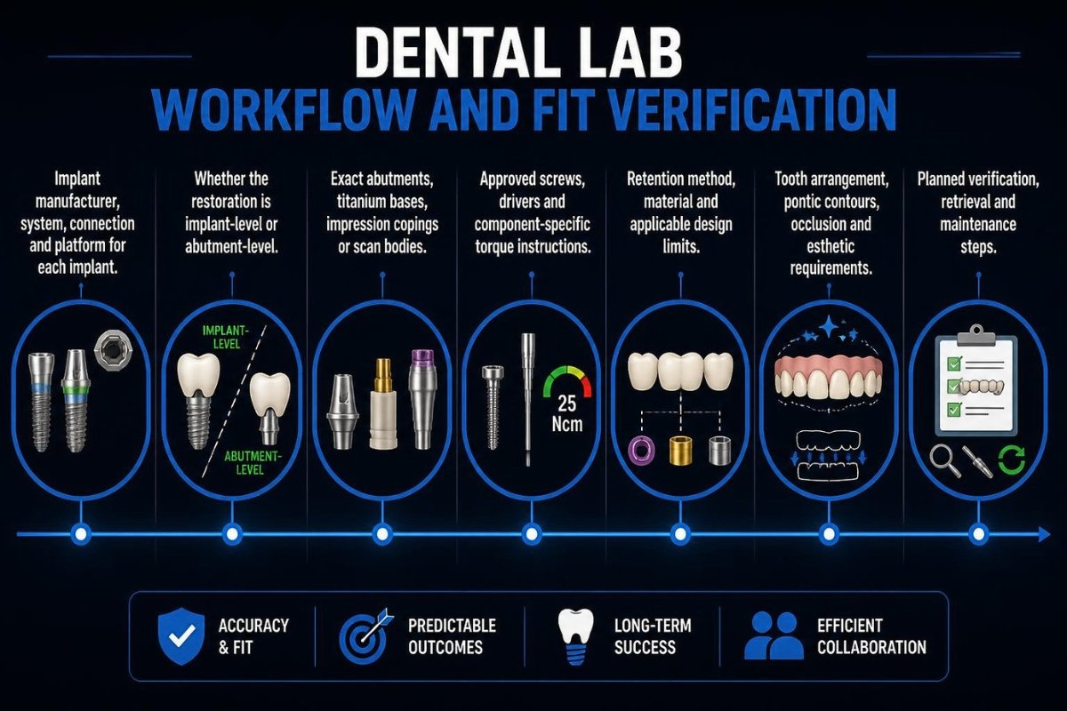
A complete prescription should identify:
- Implant manufacturer, system, connection and platform for each implant.
- Whether the restoration is implant-level or abutment-level.
- Exact abutments, titanium bases, impression copings or scan bodies.
- Approved screws, drivers and component-specific torque instructions.
- Retention method, material and applicable design limits.
- Tooth arrangement, pontic contours, occlusion and esthetic requirements.
- Planned verification, retrieval and maintenance steps.
Platform diameter alone does not establish compatibility. Third-party components require documented compatibility and appropriate regulatory consideration; an apparent fit does not prove equivalent tolerances or mechanical performance.
Conventional and digital records
Conventional workflows may use implant impression copings, master casts and verification devices. Digital workflows may use intraoral scans, implant scan bodies and, in selected multi-implant cases, photogrammetry. Hybrid workflows can combine records and verification methods.
Accuracy depends on implant span and distribution, scan-body seating, scanner and software, library selection and operator technique. Photogrammetry can help record implant positions but does not necessarily provide the tissue and occlusal information needed to complete the bridge.
A complete-looking digital model is not proof that the finished restoration will seat accurately on all supports.
How is bridge fit verified?
Clinical fit assessment may combine visual and tactile checks, appropriate radiographs, controlled screw-sequence assessments and verification devices. The team should also check for rocking, incomplete seating, interproximal interference and tissue pressure.
Screws should not be used to pull a visibly misfitting bridge into place. If seating is uncertain, the impression or scan, component selection, manufacturing process and assembly should be investigated.
Manufacturing, bonding and screw handling
Relevant manufacturing controls include milling accuracy and tool wear, metal-framework processing, zirconia shrinkage and sintering, veneering support, surface finishing and titanium-base assembly. Digital manufacturing can improve design control, but it does not replace clinical verification.
Bonding procedures must match the materials and components used. Zirconia and glass ceramics require different surface treatments. During assembly, functional implant connections and screw seats should be protected, and excess resin should be removed.
At delivery, use the exact approved screws and drivers with a maintained torque instrument. Follow manufacturer-specific torque, retightening and screw-replacement instructions. Recurrent loosening warrants investigation of fit, loading and component integrity—not repeated tightening alone.
Standards and traceability
Standards can support testing, material controls, biological evaluation, risk management and quality systems. Examples relevant to particular components or processes include ISO 6872 for dental ceramics, ISO 22674 for metallic restorative materials, ISO 14971 for medical-device risk management and ISO 13485 for quality-management systems. Their current editions and exact scopes should be verified through the official ISO catalogue.
Standards compliance is not proof that a particular bridge design will succeed clinically. A laboratory release record should also document component identity, material lots, processing, fit checks, bonding, finishing and delivery instructions.
Clinical evidence, complications and maintenance
Implant-supported bridges can remain in service for years, but prosthesis survival and complication-free function are different outcomes. Evidence should be interpreted by bridge design, material and follow-up period.
What do systematic reviews show?
A 2018 systematic review by Sailer and colleagues compared metal–ceramic and zirconia–ceramic multiple-unit fixed dental prostheses. It estimated five-year survival of 98.7% for metal–ceramic and 93.0% for zirconia–ceramic restorations in the included studies.
Those figures describe the restorations and study periods reviewed. No monolithic zirconia fixed dental prosthesis studies met that review’s inclusion criteria, so its zirconia–ceramic estimate should not be used as a survival figure for contemporary monolithic zirconia bridges.
A newer review by Lemos and colleagues addresses monolithic ceramic implant-supported single crowns and fixed partial dentures. When applying such research to a bridge case, single-crown outcomes should be separated from bridge outcomes, and short-span designs from full-arch prostheses.
Common complications
Technical complications include screw loosening or fracture, veneer chipping, framework fracture, titanium-base debonding, loss of cement retention, access-filling loss and open contacts. Biological concerns include peri-implant mucositis, peri-implantitis and inflammation associated with difficult-to-clean contours or retained cement.
The European Federation of Periodontology’s S3-level clinical practice guideline provides an evidence-based framework for preventing and treating peri-implant diseases. It reinforces why access for cleaning and professional assessment should be considered before the bridge is fabricated.
What should long-term maintenance assess?
Individualized recall visits should assess peri-implant tissues, plaque control, cleaning access, food trapping, contacts, occlusion, component stability and material integrity. Radiographic assessment may be appropriate when clinically indicated.
A fixed bridge does not automatically need removal at every recall. Persistent bleeding, suppuration, increasing probing depths, pain or mobility does require clinical assessment. Technical and biological problems can occur together and may need separate diagnosis.
Working with a dental lab partner
For dental practices using lab-to-lab services or dental lab outsourcing, a reliable handoff starts with exact implant identifiers, complete restorative records and an agreed verification plan. The receiving team should be able to trace the components and materials used and understand how the bridge can be retrieved or repaired.
XDENT LAB supports implant and removable prosthetic workflows through its lab-to-lab service from Vietnam. In an implant bridge case, its laboratory role is to follow the agreed component, material and manufacturing specifications and communicate the completed restoration’s documentation. Implant selection, clinical fit verification and ongoing patient care remain with the treating clinical team.
Key takeaway: The best implant-supported bridge is not simply the strongest framework. It is a restoration with compatible components, verified combined seating, appropriate material and geometry, cleanable contours and a realistic plan for maintenance and repair.
About XDENT LAB:
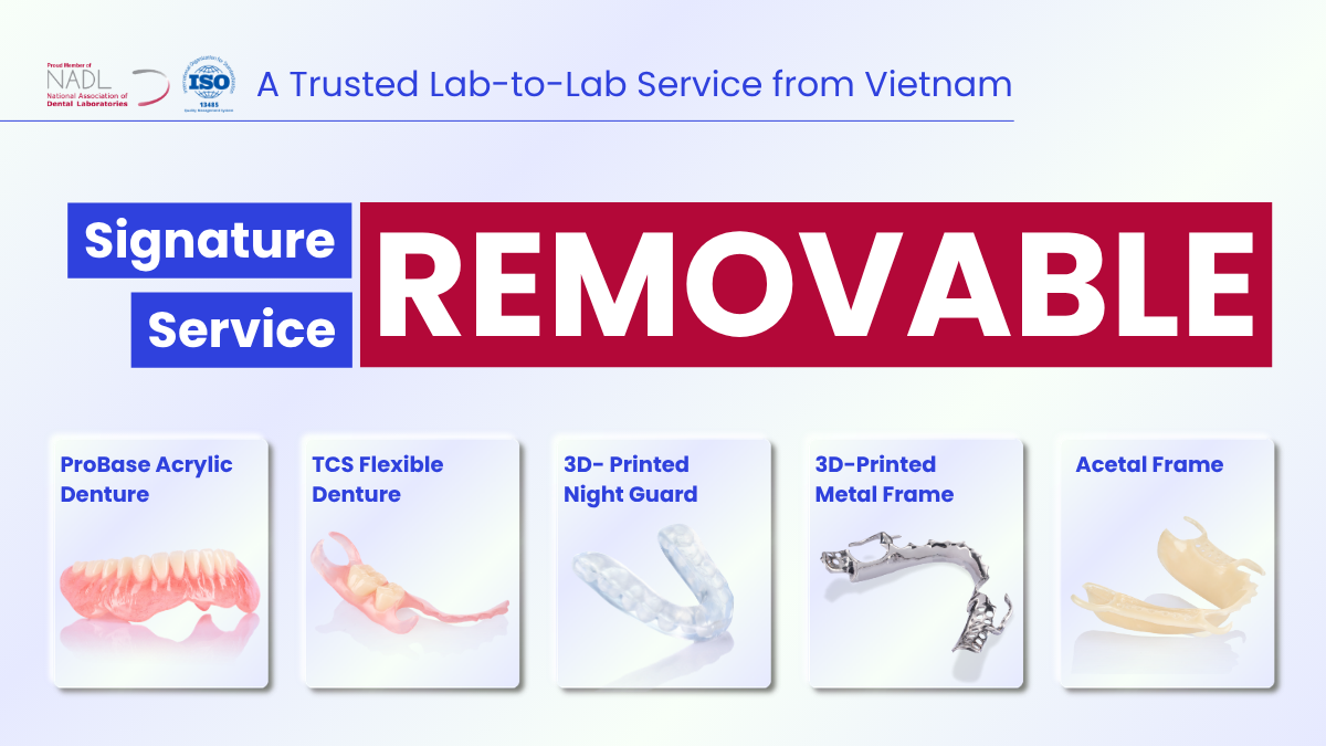
We are experts in Lab-to-Lab Full Service from Vietnam, with the signature services of Removable, meet U.S. market standards, FDA-registered, ISO 13485-certified. Founded in 2017, from local root to global reach, we scale with 2 Factories with over 100+ employees.
Our Commitments Built on “Trusted. Commitment. Quality”
- Commit to Large-Scale Manufacturing, high volume, remake rate < 1%.
- Commit to 5-Year Warranty
- Commit to Competitive Price
XDENT LAB | A Trusted Lab-to-Lab Service from Vietnam
Share this post:
Related Posts
From digital scan to final glaze, see how a full zirconia crown or bridge is designed, milled, sintered and finished, and what affects quality along the way.
A practical guide to single implant crowns, covering abutments, Ti-bases, crown materials, common complications and ongoing care.
A practical guide to implant crowns, bridges and overdentures, covering materials, component compatibility and laboratory workflow.

