
Explore the types, materials, retention methods and lab workflows for maxillofacial removable prostheses, with practical guidance for dental practices.
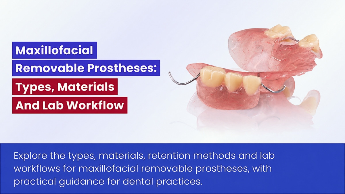
Table of contents [Show]
Maxillofacial removable prostheses are patient-specific devices that replace missing structures of the mouth or face, or help remaining structures function more effectively. They may support speech, swallowing and chewing, protect tissues, or restore facial appearance. Their design extends beyond conventional denture fabrication because anatomy, support, retention and maintenance needs differ in every case.
For dental practices, understanding these devices helps improve treatment planning and communication with the maxillofacial prosthodontist and laboratory. This guide covers the principal types, material choices and clinical-to-lab workflow.
Types of maxillofacial removable prostheses
Maxillofacial prostheses may be intraoral, extraoral or a combination of both. Patients may need them after cancer treatment, trauma, congenital conditions or other causes of tissue loss.
Intraoral prostheses
These appliances replace tissue or assist function within the mouth and adjacent spaces.
- Maxillary obturator: Separates the mouth from a defect communicating with the nasal cavity or sinus. It may also replace missing teeth.
- Speech-bulb prosthesis: Helps compensate for insufficient soft-palate tissue in selected patients.
- Palatal-lift prosthesis: Elevates a soft palate that is present but moves inadequately.
- Palatal augmentation prosthesis: Reshapes the palate to improve contact with the remaining tongue.
- Mandibular guidance prosthesis: Helps guide a deviating mandible toward a more useful closing position.
- Resection-related denture: Replaces teeth and selected tissue volume after jaw surgery.
The diagnosis determines the appliance. A speech bulb addresses insufficient tissue, while a palatal lift assists tissue that remains but functions poorly. A palatal augmentation prosthesis serves a different purpose again.
Stages of maxillary obturator treatment
Obturators may be provided at different stages of care:
- Surgical obturator: Used at or around surgery when appropriate.
- Interim obturator: Modified as the defect heals and tissues change.
- Definitive obturator: Fabricated when the anatomy and treatment plan are sufficiently stable for longer-term use.
“Definitive” does not mean permanent. Even a well-made obturator may need adjustment, repair or replacement as the patient’s needs change.
Extraoral facial prostheses
Facial prostheses replace visible structures. Their design must balance appearance with predictable placement, comfortable margins and retention the patient can manage.
- Auricular prostheses replace the external ear.
- Nasal prostheses replace part or all of the external nose.
- Orbital prostheses replace an eye and surrounding facial tissues.
- Ocular prostheses restore the visible appearance of an absent eye within a suitable socket.
- Midfacial prostheses replace larger cheek or central facial structures.
An ocular prosthesis is commonly a rigid artificial eye fitted in a socket. An orbital prosthesis replaces the eye and surrounding tissues, often with a silicone facial component. Neither restores vision.
Some patients need a combined intraoral–extraoral prosthesis. In these cases, the components must seat predictably without making cleaning or handling unnecessarily difficult.
Treatment planning, support and retention
The starting question is not simply what tissue is missing. The clinical team must also consider how the patient will place, wear, remove and maintain the prosthesis.
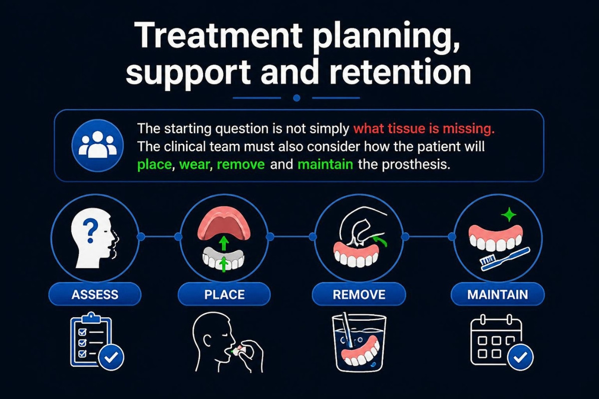
Clinical assessment
Assessment may include defect size and location, healing status, remaining teeth, available bone and soft tissue, mouth opening, saliva, speech and swallowing needs, and the patient’s dexterity and vision.
Previous or planned radiotherapy also matters. Tissue fragility, dry mouth, restricted opening and healing concerns may influence the design and follow-up plan.
Retention methods
Retention may come from remaining teeth, suitable anatomical undercuts, adhesives, spectacles, craniofacial implants or a combination of methods.
- Clasps and dental rests can support suitable intraoral prostheses when useful teeth remain.
- Precision attachments or double crowns can provide controlled retention in selected cases but require space and ongoing servicing.
- Anatomical undercuts may improve retention if engagement does not injure tissue or make removal difficult.
- Skin adhesives offer a nonsurgical option for some facial prostheses but can leave residue or irritate skin.
- Spectacles or mechanical supports may help position selected facial prostheses.
- Craniofacial implants can support repeatable placement but require site-specific planning and maintenance.
More retention is not always better. Excessive removal force can injure tissue or overload supporting structures. Results from conventional intraoral dental implants should not automatically be applied to orbital, nasal or temporal-bone sites.
Obturator or surgical reconstruction
Both approaches can be appropriate, and they may be used together. An obturator can be removed for cleaning and examination and modified as tissues change. Reconstruction may offer advantages for particular large or poorly supported defects.
A retrospective study of 113 patients reported by MD Anderson Cancer Center found comparable postoperative speech intelligibility and diet between obturator and free-flap groups overall. In that cohort, free flaps produced better functional outcomes for extensive defects involving more than half the palate. These findings inform treatment planning but do not replace individual assessment.
An obturator’s removability may facilitate examination, but it does not establish improved cancer surveillance or survival.
Materials used in maxillofacial prostheses
Material selection should reflect the finished device’s intended contact site, functional demands, processing method and maintenance needs. A material suitable for a model is not automatically suitable for a patient-contact prosthesis.
Common material choices
- PMMA acrylic resin: Used for obturators, denture bases and selected rigid components. Fit, porosity, polish and strength require attention.
- Medical-grade silicone elastomer: Commonly used for external facial prostheses. Edge durability, color stability and compatibility are important.
- Cobalt–chromium: Used for selected rigid intraoral frameworks where accurate fit and finishing are essential.
- Titanium and titanium alloys: Used for selected frameworks and implant components, subject to exact component compatibility.
- Soft liners: Used in selected interim applications but require monitoring for aging and hygiene.
- Validated printable polymers: Used for models, patterns, molds or specific approved clinical components, according to their intended use.
Material suitability must be evaluated for the complete processed device, including pigments, primers and adhesives where applicable.
Facial silicone and color stability
Silicone’s flexibility and optical properties can help reproduce skin-like contours. Natural appearance depends on translucency, regional color, texture, gloss and details such as hair or eyebrows where appropriate.
Thin edges can make a facial prosthesis less visible, but they are more vulnerable to tearing during handling or adhesive removal. The laboratory must balance marginal blending with durability.
Color can also change over time. A review of maxillofacial silicone color stability identified color deterioration as an important limitation. Its findings should not be treated as a fixed replacement schedule for every material or patient.
The clinical-to-lab workflow
Complex cases work best when clinical decisions and laboratory steps remain closely connected. The laboratory fabricates to a specialist prescription; it does not independently diagnose the defect or determine the treatment plan.
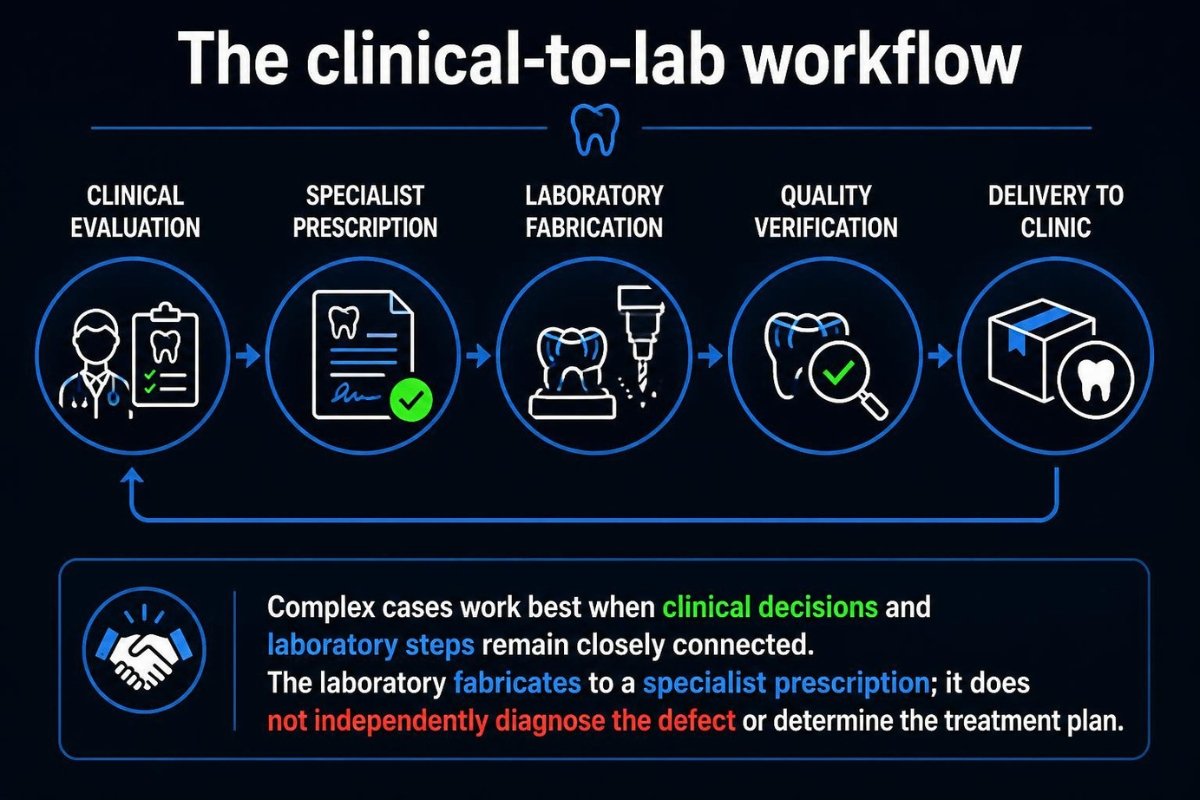
Define the functional goal and prescription
The clinical team identifies what the device must accomplish: separation of cavities, tooth replacement, speech support, facial restoration or a combination. The prescription should specify intended use, retention, materials and relevant limitations.
Capture anatomy safely
Clinical records may include impressions, facial or intraoral scans, photographs and, when justified, imaging-derived information. Complex defects can communicate with vulnerable cavities or the airway. Impressions therefore require clinical control of material flow, undercuts, fragile tissues and safe removal.
Noncontact scanning may reduce some impression-related risks, but it may not capture every deep cavity or functional tissue movement.
Design and verify the prosthesis
Conventional workflows use models, sculpted or wax patterns, clinical trials and molds. Digital workflows may add anatomical mirroring, CAD design, printed patterns or printed molds.
Printed molds and directly printed prostheses
Printing a mold for conventionally processed silicone is not the same as directly printing the final silicone prosthesis. Each approach requires its own assessment of materials, accuracy, processing and intended use. Hybrid workflows can pair digital design with clinical verification and manual characterization.
Process and document the finished device
Before release, the laboratory should verify the aspects relevant to the prescribed prosthesis, including fit, surface finish, retention-component compatibility, joins and bonding. A closed hollow-bulb obturator also needs a sound seal and a practical repair plan.
Useful documentation includes the prescription, material identities and lots, processing records, pigment formulation where applicable, fit checks, and cleaning and repair instructions.
Relevant ISO standards may address denture-base polymers, metallic dental materials, biological evaluation, risk management and quality-management systems. Current editions and scope should be checked through the official ISO catalogue. Quality-management certification does not establish the clinical performance of every individual prosthesis.
Maintenance and laboratory partnership
Delivery begins an ongoing care process. Reviews may assess tissue health, pressure areas, obturator leakage, speech and swallowing, attachment wear, implant or abutment health, surface condition, and the patient’s ability to clean and handle the device.
There is no universal replacement interval. A prosthesis may need servicing because tissues change, retention components wear, silicone margins tear or color no longer matches. New bleeding, persistent ulceration, unexplained pain or a changing lesion requires clinical assessment rather than a routine laboratory adjustment.
For practices working with an outsourced laboratory, a clear specialist prescription, complete records and a follow-up plan support consistent fabrication. XDENT LAB’s removable and implant laboratory experience can support relevant workflows, while diagnosis, treatment selection and clinical verification remain under the direction of the treating team.
Key takeaway: A successful maxillofacial removable prosthesis is more than an accurate replacement shape. It is a maintainable rehabilitation system built around the patient’s anatomy, functional goals, retention, materials and ability to use it every day.
Share this post:
Related Posts
Explore precision attachment denture types, benefits and limitations, plus key considerations for design, fabrication and maintenance in dental practices.
Explore telescopic denture materials, retention designs and lab workflows, with practical guidance on fit, fabrication and maintenance for dental practices.
Materials used in fixed prosthodontics, from zirconia and lithium disilicate to alloys, with guidance on indications, bonding, workflows, and selection.

